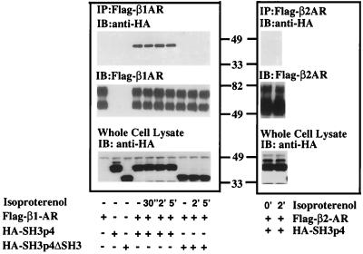
Figure 3.
Coimmunoprecipitation of HA-SH3p4 and Flag-β1-adrenergic receptor from HEK293 cells. pcDNA3 Flag-β1-AR (Left) or pcDNA3 Flag-β2-AR (Right) were transiently transfected into HEK293 cells in the absence or presence of pcDNA3 HA-SH3p4 or pcDNA3 HA-SH3p4 ΔSH3. Nonstimulated cells and cells stimulated with 10 μM isoproterenol for various periods of time were lysed in digitonin buffer. Flag-β1-AR was immunoprecipitated with M2 anti-Flag affinity gel from samples containing equal amounts of protein. Immunoprecipitates were subjected to SDS/PAGE and Western blot analysis. The blot was initially probed with anti-HA rabbit polyclonal antibody to detect HA-SH3p4 and HA-SH3p4 ΔSH3. The blot was subsequently stripped and reprobed with biotinylated-M2 anti-Flag antibody and streptavidin/horseradish peroxidase conjugates to detect Flag-βAR. Overexpression of HA-SH3p4 or HA-SH3p4ΔSH3 in whole cell lysate also was monitored. The experiment was repeated at least three times with similar results. The diffuse doublet of both Flag-β1-AR and Flag-β2-AR in the middle panel likely represents differentially glycosylated forms of these receptors.

