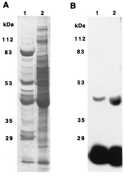
Figure 4.
Comparison of 43-kDa protein in Blue Sepharose and S200-B fractions by SDS/PAGE. Blue Sepharose (lanes 1) and S200-B (lanes 2) fractions were incubated for 15 min at room temperature with 250 nM [32P]NDPK. After these incubations the samples were separated on SDS/PAGE. The gel was stained with Coomassie blue in 10% acetic acid and 25% isopropanol for 1.5 hr, destained in 10% acetic acid for 2 hr, and dried. A is the Coomassie brilliant blue-stained gel, and B is the autoradiogram of this gel.

