Figure 3.
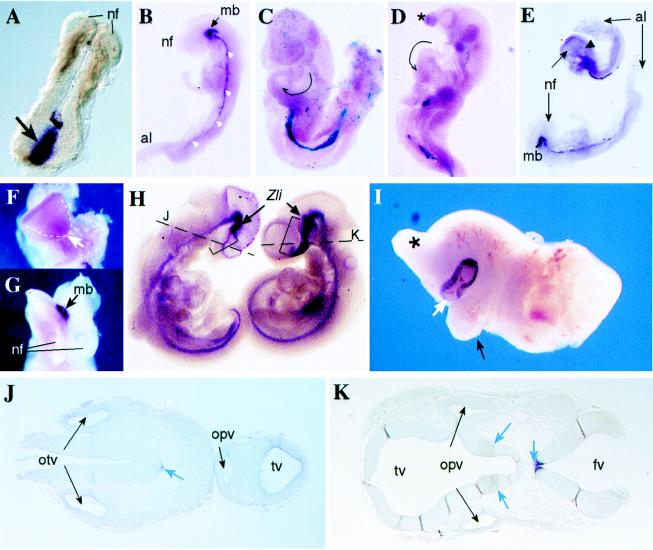
Rescue of Smad2−/− phenotype and chimeric embryo phenotypes. (A) Brachyury in situ hybridization in a day 7.5 Smad2−/− embryo recovered from a tetraploid ⇔ Smad2−/− chimera. A strong staining of this mesoderm marker is observed in the primitive streak area (arrow). The formation of the neural folds (nf) in the anterior part of the embryo is also evident. (B) Shh in situ hybridization in a day 8.5 Smad2−/− rescued embryo. Staining for this marker is observed in the axial mesoderm (white arrowheads), as well as in the ventral midline of the midbrain (mb). The neural folds (nf, anterior) and the allantois (al, posterior) are indicated. (C) X-gal staining in a day 9.5 tetraploid ROSA26 ⇔ Smad2−/− chimera (viewed from its left side) showing that the embryo is formed almost exclusively by Smad2−/− cells. This particular embryo showed the highest contribution of wild-type tetraploid cells to the embryo (blue cells in the primitive endoderm, the allantois region, and head mesenchyme) and yet the embryo showed turning in clockwise direction (tailbud to the left) and bulbus cordis of the heart looping to the left of the midline (arrow). In normal embryos, the position of the bulbus cordis is at the right of the midline. (D) X-gal staining in a day 10.5 tetraploid ROSA26 ⇔ Smad2−/− chimera viewed from the left showing absence of turning (posterior part U-shaped) and abnormal head formation (∗), but normal turning of the cardiac tube (arrow). Some blue wild-type cells are observed only in the region of the primitive endoderm. (E) Shh in situ hybridization of two rescued embryos at day 8.5. Although the embryo in the bottom shows normal shh expression along the midline and midbrain (mb), in the upper embryo the expression of shh extends just to the hindbrain region (arrowhead). nf, neural folds; al, allantois. (F) Frontal view of the upper embryo shown in E, lacking normal midline separation at the anterior neural folds (arrow). (G) Frontal view of the bottom embryo shown in E exhibiting normal separation of the neural folds (nf). The expression of shh in the ventral midbrain (mb) is indicated. (H) Shh in situ hybridization in a tetraploid ⇔ Smad2−/− chimera (Left) and a wild-type (Right) embryo. The staining in the region of diencephalon and telencephalon (brackets) anterior to the zona limitans (Zli) is absent in the mutant cyclops embryo (see below). Moreover, in the Smad2-deficient embryo, the tip of the tail is positioned to the left, in contrast with the rightwards direction shown by the wild-type embryo. (I) Anterior fragment of a diploid ⇔ Smad2−/− chimera recovered at day 13.5 showing cyclopia. A proboscis (∗) and a protruding remnant of the right branchial arch (black arrow) are observed. The large contribution of mutant ES cells to this chimera is revealed by the strong pigmentation of the single eye (white arrow). (J) Transverse section of the mutant embryo shown in D (at the level of the broken line) shows a single telencephalic vesicle (tv) and a single optic vesicle (opv). A weak Shh staining in the floor plate of the neural tube and notochord (white arrow) and both otic vesicles (otv) are indicated. (K) Transverse section of the wild-type embryo shown in D (at the level of the broken line) shows normal bilobar telencephalic vesicles (tv) and two optic vesicles (opv). Normal Shh expression in the floor plate and ventral diencephalon is shown (blue arrows). The fourth ventricle (fv) is indicated.

