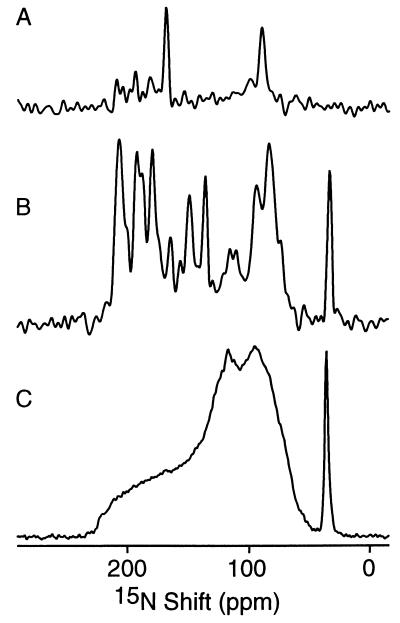
Figure 1.
One-dimensional 15N chemical shift NMR spectra of 15N-labeled fd coat protein in phospholipid bilayers. (A) Selectively 15N Leu-labeled protein in oriented bilayers. (B) Uniformly 15N-labeled protein in oriented bilayers. (C) Uniformly 15N-labeled protein in unoriented bilayer vesicles. Two hundred and fifty-six data points were acquired using CPMOIST cross polarization with a mix time of 1 ms and a recycle delay of 3 sec.

