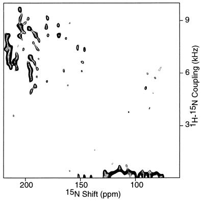
Figure 2.
Two-dimensional 1H–15N dipolar coupling/15N chemical shift PISEMA spectrum of an oriented sample of uniformly 15N-labeled fd coat protein in phospholipid bilayers. Two hundred and fifty-six transients were acquired for each of 64 t1 values incremented by 40.8 μsec. The cross-polarization mix time was 1 msec and the recycle delay was 3 sec.

