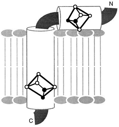
Figure 5.
The orientations of the peptide planes for Leu-14 and Leu-41 of fd coat protein in phospholipid bilayers, as determined from the data in Fig. 3 F and C. The planes are placed in the context of the structure determined by solution NMR spectroscopy of this membrane protein in micelles, which has a hydrophobic trans-membrane helix and an amphipathic in-plane helix.

