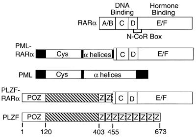
Figure 1.
Schematic representations of the RARα, PML, PML-RARα, PLZF, and PLZF-RARα proteins. Proteins are depicted from N to C terminus. Sequences derived from RARα are subdivided into domains A to F, as previously described, with the receptor regions involved in DNA and hormone binding indicated above the schematic, and the receptor N-CoR box, necessary for corepressor association, indicated below (1–6). Putative structural motifs identified within PML or PLZF-derived sequences include a cysteine-rich RING finger/B-box motif (Cys), an α-helical/coiled-coil domain (α-helices), a POZ domain (37), and zinc-finger motifs (Z). Codon positions within PLZF are numbered from the N terminus.

