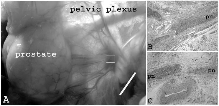
Fig. 3.
Gross and microscopic view of the pelvic plexus and pelvic nerve one week post PDT. (A) Hyperemia lesions induced by 1 mg/kg and 50 J/cm2. The lesion size was smaller than the size of irradiated area (i.e. 1 cm). (Bar = 1 cm) (B) Microscopic view of the marked area of the previous photo. H&E staining of longitudinal section of pelvic plexus showed the segmental endoneurial hemorrhage of the pelvic nerve fascicles (PN). Degeneration nerve fibers and necrotic neurons were present. The adjacent connective tissue showed areas of hemorrhage and fibrosis, regional accumulations of inflammation cells in varying proportions. (C). Microscopic view of the control (no drug, no light). (×20)

