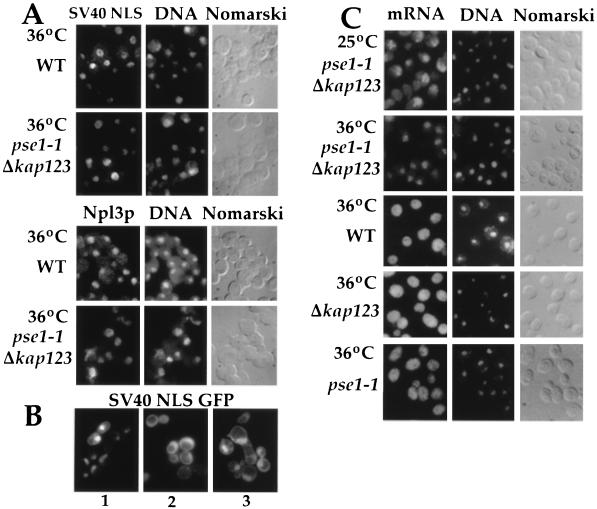
Figure 3.
Protein and mRNA transport in pse1–1 Δkap123 mutants. (A) Cells were grown in selective media and the SV40 NLS invertase was expressed under the control of the ADH1 promotor and visualized by immunofluorescence using an invertase specific antibody (Upper). Npl3p was visualized by immunofluorescence using a Npl3p specific antibody (Lower). (B) pse1–1 Δkap123 cells expressing NLS-GFP were grown at 25°C (panel 1), washed into glucose-free media with 10 mM sodium azide and 2-deoxy-d-glucose for 45 min (panel 2), and then shifted to 36°C for 30 min and then washed into prewarmed glucose-containing media and incubated for 15 min at 36°C (panel 3). (C) Poly(A)+ RNA was detected using an in situ hybridization assay with an oligo dT(50) probe (37).

