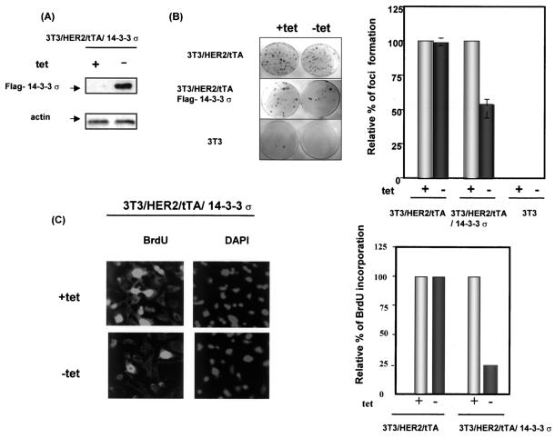
FIG. 6.
Tetracycline-regulated 14-3-3σ inhibited transformation phenotype and mitogenic signals of HER2/neu-overexpressing cells. (A) Tetracycline-inducible expression of 14-3-3σ. To examine the expression of 14-3-3σ, the culture medium of 3T3/HER2/tTA/14-3-3σ was switched to medium alone or medium containing 2 μg of tetracycline (tet) per ml. The Flag-14-3-3σ expressed in the absence of tetracycline was probed by immunoblotting with anti-Flag antibody. The levels of actin were used as an equal loading control. (B) Focus formation assay. NIH 3T3 (3T3), 3T3/HER2/tTA, and 3T3/HER2/tTA/Flag-14-3-3σ cells were subjected to a microfocus formation assay in the presence (+) or absence (−) of 2 μg of tetracycline per ml. Foci were counted in the presence and absence of tetracycline. The number of foci from 3T3/HER2/tTA and 3T3/HER2//tTA/14-3-3σ cells treated with tetracycline was set at 100%. The relative focus formation from cells cultured in the absence of tetracycline is shown as a bar graph. Bars represent standard deviations. (C) Tetracycline-regulated 14-3-3σ expression blocked cell cycle entry into S phase in HER2/neu-overexpressing cells. Bromodeoxyuridine (BrdU) incorporation assays were conducted after cells were cultured in the presence or absence of tetracycline for 20 h. Incorporation of bromodeoxyuridine was examined under a fluorescence microscope with fluorescein isothiocyanate-conjugated antibromodeoxyuridine. DAPI staining indicated the location of nuclei. A total of 300 cells were counted for bromodeoxyuridine staining in each condition. The number of bromodeoxyuridine-positive cells from 3T3/HER2/tTA and 3T3/HER2//tTA/14-3-3σ cells cultured in tetracycline-containing medium was set at 100%. The relative percentage of bromodeoxyuridine-positive cells in cells cultured in the absence of tetracycline is presented. Data shown are from a typical experiment conducted in triplicate.

