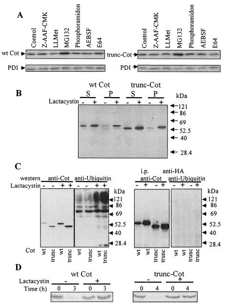
FIG. 3.
Levels of wt Cot and trunc-Cot expression after treatment of HEK293 transfected cells with different protease inhibitors. (A) Total extracts of HEK293 cells 14.5 h after transfection with 10 μg of pcINEO HA-wt Cot or 10 μg of pcINEO HA-trunc-Cot were further incubated for 7.5 h in the presence of different protease inhibitors (10 μM Z-AAF-CMK, a tripeptidyl peptidase inhibitor; 20 μM LLMet, a calpain II inhibitor; 20 μM MG132, a proteasome inhibitor; 20 μM phosporamidon, a metalloprotease inhibitor; 1 mM prefabloc AEBSF, a serine protease inhibitor; or 20 μM E64, a cysteine protease inhibitor) as indicated in Materials and Methods. Total extracts were analyzed by Western blotting using anti-Cot (Calbiochem) and anti-PDI antibodies. The figure shows representative results of one of the three experiments performed. (B) Cells transfected as indicated for panel A and incubated in the presence or absence of 10 μM lactacystin were lysed in lysis buffer, and both the 1% NP-40-soluble and -insoluble fractions were analyzed by Western blotting with an anti-Cot antibody (Calbiochem). (C) Total extracts of cells 14.5 h after transfection with 5 μg of pcDNA3.1 HA-wt Cot or 5 μg of pcDNA3.1 HA-trunc-Cot together with 5 μg of pCMV-His-ubiquitin, which were further incubated for 7.5 h in the presence or absence of 10 μM lactacystin, were analyzed by Western blotting with anti-Cot antibody (Calbiochem) or with antiubiquitin antibody. Both wt Cot and trunc-Cot were immunoprecipitated from the same transfected cells and analyzed by Western blotting with antiubiquitin antibody and anti-Cot antibody. The figure shows representative results of one of the three experiments performed. (D) Accumulation of newly synthesized wt Cot and trunc-Cot after lactacystin (10 μM) incubation. 35S-labeled wt Cot and trunc-Cot were immunoprecipitated from HEK293 cells transfected with 10 μg of pcINEO HA-wt Cot or 10 μg of pcINEO HA-trunc-Cot after a 20-min pulse and 3-h chase (wt Cot) or 4-h chase (trunc-Cot) in the presence or absence of 10 μM lactacystin. The proteins were resolved by SDS-PAGE and visualized by autoradiography. The figure shows representative results of one of the three experiments performed.

