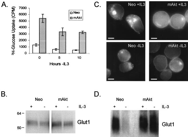
FIG. 3.
Constitutively active Akt promotes glucose uptake not by regulating Glut1 protein levels but by promoting Glut1 cell surface localization. (A) Control (Neo) and mAkt-expressing IL-3-dependent cells were cultured in the presence of IL-3 or in the absence of IL-3 for 5 or 10 h. The cells were then harvested, and their ability to uptake the nonhydrolyzable glucose analog 2-DOG was measured. The values shown are the means of the results for triplicate samples; error bars indicate standard deviations. (B) Control and mAkt cells were analyzed by Western blot for expression of the glucose transporter Glut1, either in the presence of IL-3 or after 10 h of withdrawal from IL-3. Each lane was loaded with 10 μg of protein. (C) Control (Neo) and mAkt cells were stained by immunofluorescence for Glut1 localization in the presence of IL-3 and after 10 h in the absence of IL-3. Each micrograph is individually contrasted to allow the best visualization of Glut1 localization. The bars represent 5 μm. (D) Plasma membranes were isolated from control and mAkt-expressing cells in the presence of IL-3 or after 12 h in the absence of IL-3, and 10 μg of protein was analyzed by Western blot for cell surface expression of Glut1.

