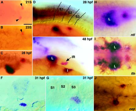
FIG. 5.
Colocalization of prox1 and ff1b at interrenal tissue in wild-type embryo and midline mutants. (A to E) Dorsal (A to C) and lateral, (D and E) view of wild-type embryos after two-color in situ hybridization with prox1 (blue) and ff1b (red) at the 21-somite stage (B), the 23-somite stage (B), 28 hpf (C), and 48 hpf (D and E). The ff1b-expressing interrenal primordium at the 21- and 23-somite stages is denoted by an arrowhead. Boundaries defining the first three somites are indicated in panel D. (F) A cross-section of a 31-hpf embryo at the midlevel of the interrenal primordium after double staining of prox1 (blue) and ff1b (red). (G) Lateral view of a 31-hpf embryo showing 3β-Hsd enzymatic activity (histochemistry) and prox1 expression (in situ hybridization). The yolk was manually removed. (H to J) Mutants with defects in midline specification including no tail (ntl) (H), floating head (flh) (I), and one-eyed pinhead (oep) (J) after double in situ hybridization of prox1 (blue) and ff1b (red). All panels show the trunk of the embryo and, except for panel F, are oriented with the anterior to the left. L, liver; S1 to S3, somites 1 to 3. Bar, 50 μm.

