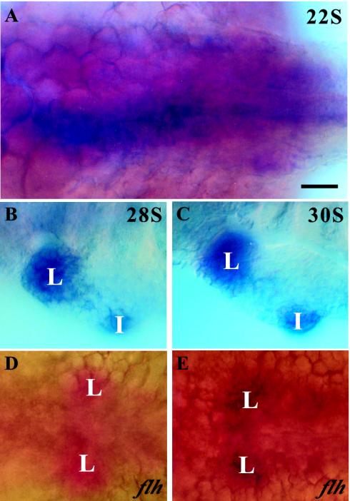
FIG. 6.
Colocalization of ff1a and prox1 in the liver primordium. Wild-type and flh mutant embryos were double labeled with ff1a (blue) and prox1 (red). (A) At the 22-somite stage, ff1a but not prox1 was detected at various regions of the posterior endoderm. (B and C) From the 28-somite stage (B) to the 30-somite stage (C), ff1a was expressed at both hepatic and intestinal primordia while prox1 colocalized with ff1a at the hepatic but not the intestinal primordium. The fast red staining of prox1 was difficult to distinguish from the strong intensity of ff1a staining. (D and E) A sequential double in situ hybridization was performed on the flh mutant, initially with prox1 (red) (D) and then with ff1a (blue) (E), with both stains delineating the symmetric hepatic primordia across the midline. L, liver; I, intestine. Bar, 50 μm.

