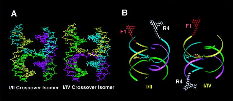
Figure 2.
Molecular models of 32 base pair immobilized HJs. (A) Full atom representation of J1 showing the geometrical relationships between the crossover isomers. In the I/II crossover isomer, arm I is stacked on arm II, and arm III on arm IV. The alternate I/IV crossover isomer has arm I stacked on arm IV, and arm II on arm III. Strand 1 is yellow, strand 2 is green, strand 3 is magenta, and strand 4 is cyan. (B) Ribbon diagrams showing the location of the FRET and NMR probes used in these studies. The F1R4 dye-labeled junction for FRET experiments is shown in the two crossover isomers, with fluorescein attached to arm I (F1) in orange and tetramethylrhodamine attached to arm IV (R4) in white. The full molecular structure of the base pair containing the labeled T9 residue for the NMR experiments is also shown, with the 15N-enriched nitrogen atom in red.

