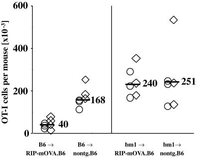
Figure 6.
Cross-presentation of high-dose OVA is essential for the deletion of OT-I cells. RIP-mOVA mice and nontransgenic littermates on a B6 (H-2b) background were lethally irradiated with 900 cGy and then reconstituted with either B6 or bm1 (H-2bm1) bone marrow (3). Eight weeks later, 9 × 10⁶ Rag-1-deficient OT-I cells were transferred into these mice. After another 6 wk, the total number of OT-I cells remaining in the spleen and the LNs of the recipient mice was determined by flow cytometry (4). The bars indicate the average number of OT-I cells found in the respective groups. These data were obtained in two separate experiments identified by ○ or ⋄. It should be noted that in earlier publications we examined deletion using B6→bm1 RIP-mOVA chimeras and justified the necessity to use such chimeras because normal B6 RIP-mOVA mice became diabetic when transferred with the large doses of cells (i.e., 5 × 10⁶ cells). For an as yet undetermined reason, B6→B6 RIP-mOVA chimeras are less sensitive to induction of diabetes than normal B6 RIP-mOVA mice and can often be transferred with 5 × 10⁶ OT-I cells without leading to diabetes. This was the case for mice used in this experiment.

