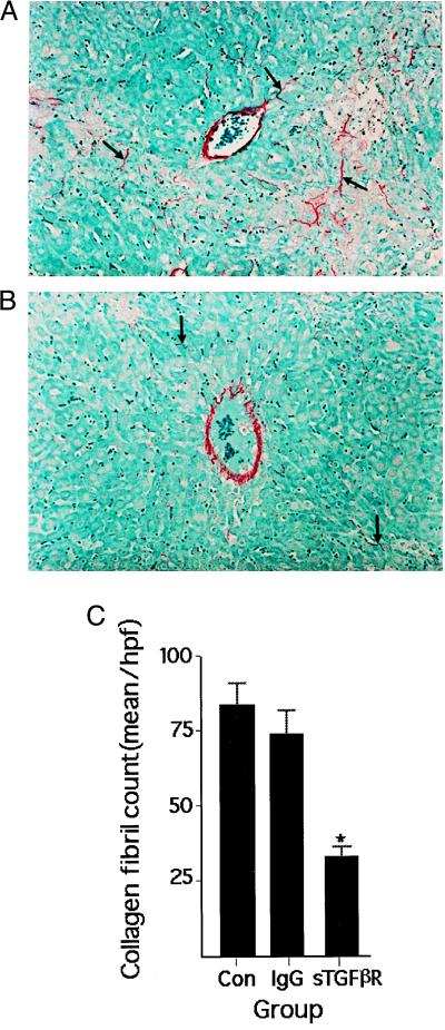
Figure 6.
Collagen in histological sections of rat liver stained with sirius red. Representative photomicrographs of liver obtained from animals treated with either control IgG (IgG) (A) or sTGF-βR (B). Rats underwent bile duct ligation on day 0 and were infused with the sTGF-βR 4 days later. Liver was harvested on day 8. In sTGF-βR-treated animals, lobular collagen is reduced. (C) Quantitation of collagen deposition in similarly treated rats. Collagen fibrils were counted in 10 high-power fields per section. Values represent the mean ± SEM of at least four separate experiments. ∗, P < 0.0002 vs. Con or IgG-treated rats. There was no difference in collagen I deposition between control and IgG-treated animals.

