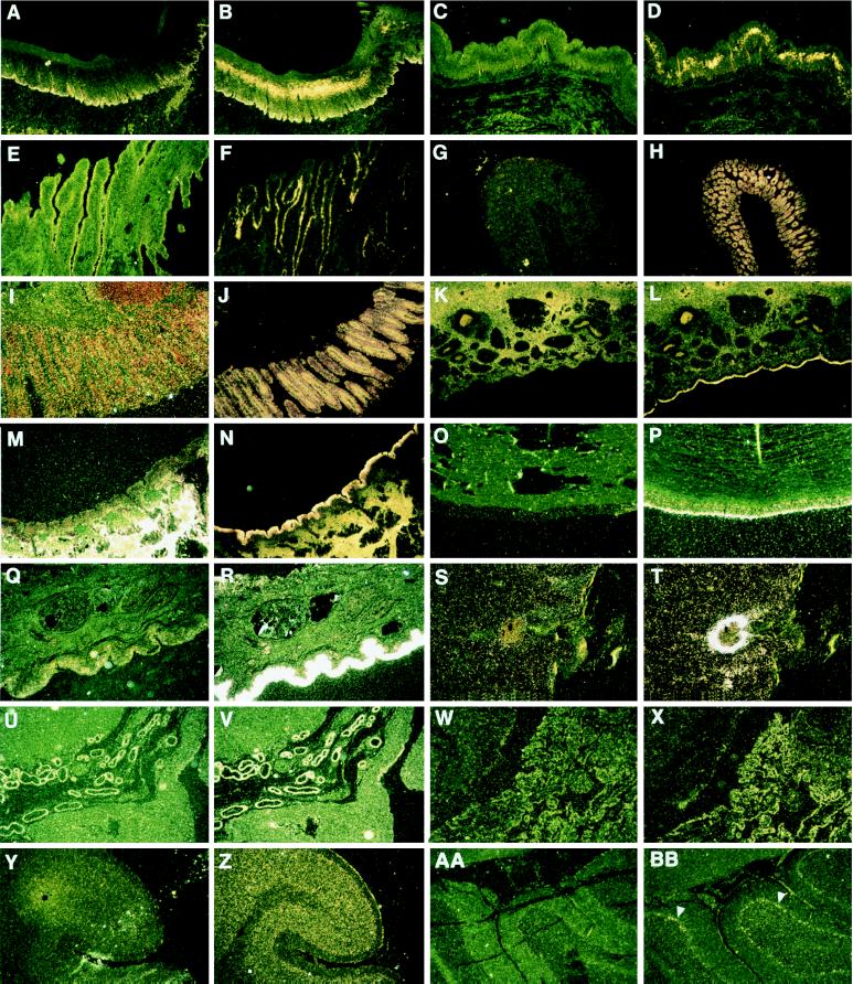
Figure 1.
In situ hybridization of LAP probe to normal and abnormal bovine tissues. The first panel of each pair represents tissue probed with the sense negative control; the second is hybridized with the antisense LAP probe. Palate (A, B), esophagus (C, D), reticulum (stomach) (E, F), colon (G, H), rectum (I, J), nares (K, L), trachea (M, N), conjunctiva (O, P), inflamed skin (Q, R), dermal abscess (S, T), placenta (embryonic epithelium) (U, V), choroid plexus (W, X), cortex of brain (granular layer and meninges) (Y, Z), and Purkinje cells (arrows: AA, BB). (Magnification ×20.)

