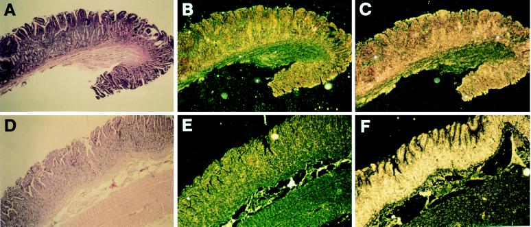
Figure 2.
Expression of β-defensins in ileum of paratuberculosis-infected cattle. (A–C) Serial sections of a portion of the ileum of a representative paratuberculosis-free animal. (D–F) Similar sections from a representative paratuberculosis-infected animal. (A and D) Hematoxylin and eosin-stained sections. (B and E) In situ hybridization with sense LAP probe. (C and F) In situ hybridization with antisense LAP probe. Uniform hybridization is seen overlying the lumenal epithelium. (Magnification ×30.)

