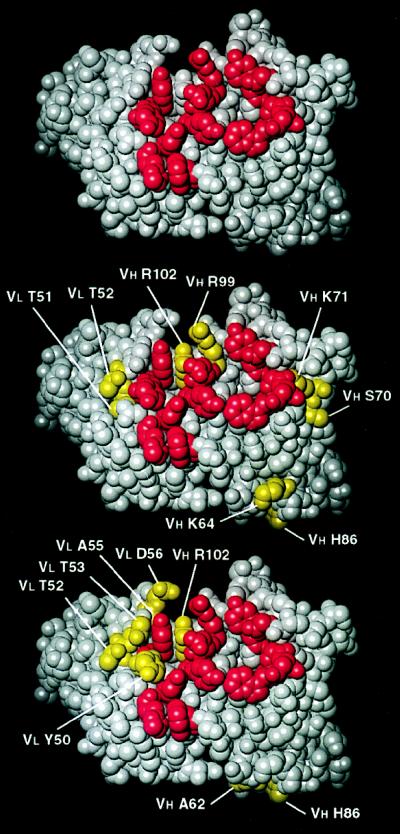
Figure 5.
Three-dimensional models of the Ab1 D1.3 (10, 11) (Top), the anti-anti-Id AF14 (Middle), and the AF52 (Bottom). AF14 and AF52 were modeled based on their close sequence homology with D1.3. Residues in red are those that, in D1.3, contact both the antigen HEL and the anti-Id, E5.2. In this front view of the combining site, residues of AF14 and AF52 (in yellow) that differ from those of D1.3 are labeled.

