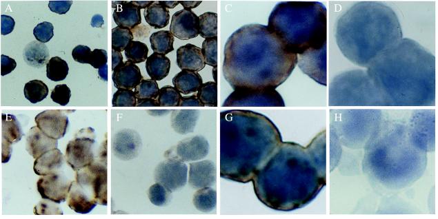
Figure 3.
(A) HCV ISH of HCV-inoculated G4 cells that were not up-regulated. Dark brown cytoplasmic staining indicates the presence of the virion form of HCV; only ≈30% of the cells are weakly positive. (B) By contrast, HCV was present in most cells in cultures in which the LDL receptor was up-regulated before HCV exposure. Original magnification for A and B, ×500. (C) High-power view of HCV-infected G4 cells with up-regulated LDL receptor as in B. (D) G4 cells prepared as in B and C were pretreated with anti-LDL receptor antibody. Blocking of the receptor by the antibodies decreased the uptake of the virus below the detection limit of ISH. Control antisera (see text) did not have such blocking effect (not shown). Original magnification for C and D, ×1,250. (E) Uptake of HCV by Hep G2 hepatoma cell line is shown by ISH. (F) Blocking the LDL receptor with its specific antibody completely prevented the endocytosis of the virus, as demonstrated by ISH in E. Original magnification for E and F, ×500. (G) Incubation of Daudi cells with HCV-positive serum resulted in positive staining by ISH. (H) Pretreatment of Daudi cells with 2 μM PAO completely inhibited the endocytosis of HCV, as illustrated in G. Original magnification for G and H, ×1250.

