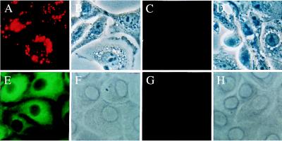
Figure 5.
(A) Intense uptake of DiI-LDL by a monolayer of the MDBK cells. (B) Phase contrast microscopy of the same field as A. (C) The lack of endocytosis of DiI-LDL was demonstrated in the CRIB cell line that is resistant to BVDV infection. (D) Same field as in C with phase contrast. (E) Demonstration of the infection of MDBK cells with the NY-1 noncytopathic strain of BVDV after 72 hours of incubation by immunofluorescence using anti-BVDV (green fluorescent staining). (F) Phase contrast picture of E. (G) No BVDV was demonstrated by immunofluorescence in the BVDV-resistant CRIB cells that have been incubated with the virus. Conditions were as in E–F. (H) Phase contrast for G. Original magnification for A–H, ×500.

