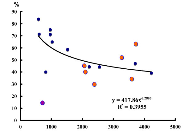
Figure 3.
Comparison of two different vector libraries in the ratio of unigenes. The unigene rate against the number of ESTs analyzed was plotted for 18 different tissue libraries. Vertical axis, percentage (number of unigenes/the number of ESTs analyzed); horizontal axis, the number of ESTs analyzed in each library. Blue, the libraries of lambda-ZAP II; orange, the libraries of pGEM. The CLOBB algorithm was used for unigene calculation. The HA (female head) library, shown in violet, was excluded from calculation of the regression curve because this library showed an extremely low unigene rate against the number of ESTs (111/777 = 0.143).

