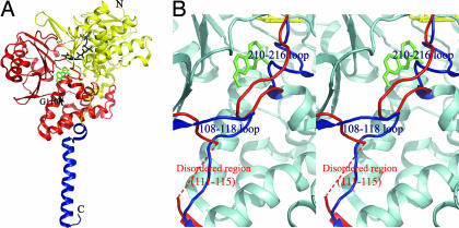
Fig. 1.
The structure of human MAOA and the comparison with the early known structure. (A) The overall structure drawn in ribbon mode. N, N terminus; C, C terminus. The structure can be divided into two domains, extra-membrane domain (shown in yellow and red) and membrane binding domain (shown in blue). The extra-membrane domain was further divided as two regions, FAD binding region (yellow) and substrate/inhibitor binding region (red). FAD (black) and harmine (green) molecules are shown as stick models. The black arrow indicates the position of G110, a residue at which we introduced mutations. (B) Stereoview of the superposed structures of human MAOA and the early published human MAOA. The identical parts between the two structures are shown in cyan. The different folds at loops 108–118 and 210–216 in the two structures are shown in blue (our structure) and red (early known structure). The fragment of A111-V115 in the early structure is disordered and not visible. Pictures were generated by PyMOL (26).

