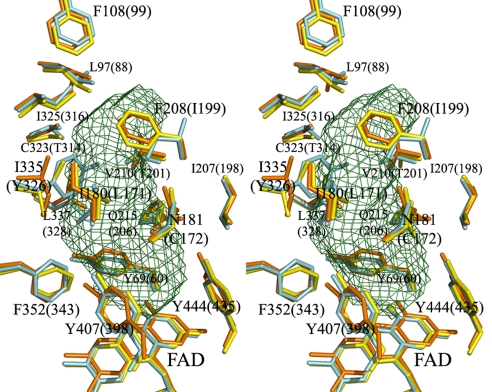
Fig. 5.
Stereoview of the substrate/inhibitor binding sites of human MAOA, human MAOB, and rat MAOA. Residues of human MAOA are shown in yellow, rat MAOA in orange, and human MAOB in cyan. The residues that are important in forming the substrate/inhibitor cavity are labeled. The residue numbering is according to the residue positions in human MAOA, which are the same as in rat MAOA. The residue numbers of human MAOB are shown in parentheses. Two residues, I199 of human MAOB and I335 of human or rat MAOA, are present as different rotamers in different complexes. The cavity was calculated by VOIDOO (13) with a 1.57-Å radius probe. These models were generated in PyMOL (26) (rmsd was 0.545 Å for human MAOB and 0.612 Å for rat MAOA).

