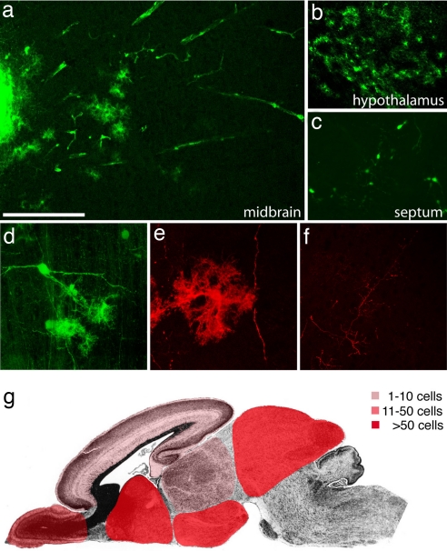
Fig. 2.
Extensive migration and differentiation of iPS cell-derived neural precursor cells in the embryonic brain. (a) Transplanted cells form an intraventricular cluster (left part of the image) and migrate extensively into the tectum 4 weeks after transplantation into the lateral brain ventricles of E13.5 mouse embryos. (b) A high density of integrated astrocyte-like cells in the hypothalamus. (c) Complex neuronal morphologies of GFP-positive cells in the septum. (d) Confocal reconstruction of grafted GFP-fluorescent cells in the tectum with neuronal and glial morphologies. (e) GFP immunofluorescence and confocal reconstruction identifies an astrocytic cell and a long neuronal process. (f) Similarly, GFP-immunoreactive, fine neuronal processes are crisply delineated. (g) Schematic representation of the main integration sites of iPS cell-derived neurons and glia. Color codes represent different ranges of cell numbers. Shown are the highest incorporation density across all analyzed brains. See Table 1 for more details. [Scale bar in a: 200 μm (a–c), 100 μm (d and f), and 50μ (e).]

