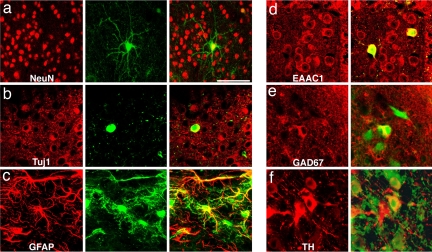
Fig. 3.
Transplanted cells express neuronal and glial markers. (a) Confocal reconstruction of a GFP-positive cell in the midbrain (green) expressing NeuN (red) 4 weeks after intrauterine transplantation. (b) Another transplanted neuron (green) expresses cytoplasmatic β-III-tubulin (red) as shown in this confocal section. (c) Other cells can be colabeled with GFAP antibodies (red). (d) Both host neurons (red only) and transplanted cells (yellow) express the glutamate transporter EAAC1. (e) Soma of grafted cells (green) can be labeled with antibodies against GAD67 (red). (f) TH-immunoreactivity (red) can be found in both host and grafted neurons (green). [Scale bar in a: 100 μm (a–c) and 50 μm (d–f).]

