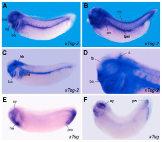
Fig. 3. Whole-mount in situ hybridization analysis of xTsg-2 and xTsg expression.

(A,C)xTsg-2 expression at stage 27 and (B,D) stage 33 embryos showing expression in the branchial arches (ba), neural crest (nc), lateral plate mesoderm (lpm) and head mesenchyme (hm). Embryos in C and D were made transparent in Murray’s Clearing Solution (2:1 benzyl benzoate: benzyl alcohol) to visualize the expression of xTsg-2 localized to the midbrain-hindbrain border, the isthmus (is). D is a close up of the embryo in B. (E) Stage 27 and (F) stage 33 embryos stained for xTsg and cleared in Murray’s solution reveal expression in the dorsal eye (ey), heart anlage (he) and proctodeum (pro), as well as in the posterior wall (pw) of the tailbud. cg, cement gland; fb, forebrain; hb, hindbrain; pn, pronephros.
