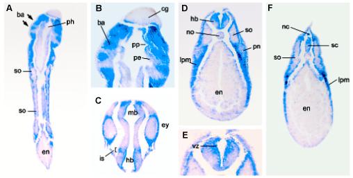
Fig. 4. xTsg-2 is expressed in the mesenchyme of the branchial arches, lateral plate and other tissues.

Histological sections of whole-mount in situ hybridization. (A) Coronal section of an embryo at stage 33 showing expression of xTsg-2 in the branchial arches (ba), but not in the pharynx (ph). (B) Close up of (A) indicating the expression of xTsg-2 is restricted to the branchial arches (ba), but devoid of detectable expression in the pharyngeal pouches (pp) consisting of pharyngeal endoderm (pe) as well as the cement gland (cg). (C) xTsg-2 expression is observed at low amounts in the eye (ey), but strong in the surrounding head mesenchyme. The isthmus (is) is stained in this coronal section. (D) Transverse section of a stage 33 embryo at the level of the trunk showing xTsg-2 expression in the lateral plate mesoderm (lpm) and hindbrain (hb) and low levels in the pronephros (pn). The somites (so), endoderm (en) and notochord (no) are not stained. (E) Close up showing xTsg-2 expression in the ventricular (proliferative) zone (vz) of the hindbrain. (F) Transverse section showing xTsg-2 expression in the dorsal fin neural crest cells and dorsal spinal cord (sc).
