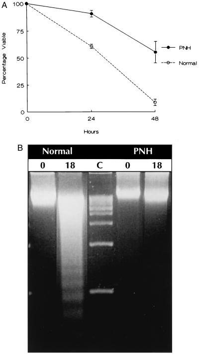
Figure 1.
(A) Survival of PNH and normal peripheral blood granulocytes in serum-free medium. Cell viability was determined by trypan blue dye exclusion. Each data point indicating percent survival represents the ratio of the absolute viable cell number at the time of assay and the number of viable cells at the start of each experiment. The data are the mean ± SEM of four patients with PNH and five normals. (B) Gel electrophoresis for detection of oligonucleosomal DNA fragments at time 0 and 18 hr after culture of normal and PNH granulocytes in serum-free medium. Lane C depicts 1-kb DNA size markers.

