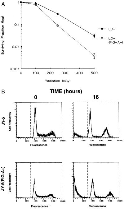
Figure 5.
Relative sensitivity of PNH cell lines to irradiation. (A) The surviving fraction from LD− and LD− (PIG-A+) cells represents the number of clonogenic colonies after treatment compared with the corresponding untreated controls. Each data point represents the mean ± SEM of three separate experiments. (B) Since JY-5 cells were unable to be cloned in semi-solid medium, flow cytometric analysis was used to assess apoptosis after 500 cGy of ionizing radiation. Propidium iodide staining of the JY-5 cells for DNA content revealed 10.5% ± 2.3 subdiploid DNA 16 hr after radiation. In contrast, JY-5 (PIG-A+) cells revealed 31.3% ± 2.9 subdiploid DNA after identical conditions. A representative example from one of the three experiments is shown.

