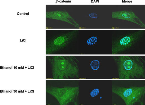
Fig. 4.
Human bone marrow cells were stained with β-catenin antibody (green) and the cells were costained with DAPI to make the cell nuclei (blue). Immunofluorescent images were observed with a confocal microscope. After the treatments with ethanol, immunofluorescence staining showed β-catenin–FITC complexes decreased in the nucleus. The results support our second hypothesis.

