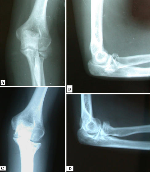
Fig. 2A–D.
(A) An anteroposterior radiograph shows a Mason Type III fracture of the radial head. (B) The lateral radiograph of the same patient shows the fracture’s comminution. (C) A postoperative radiograph obtained at the final followup shows successful reconstruction of the radial head. (D) A postoperative lateral radiograph shows fracture union and satisfactory alignment of the radiohumeral joint.

