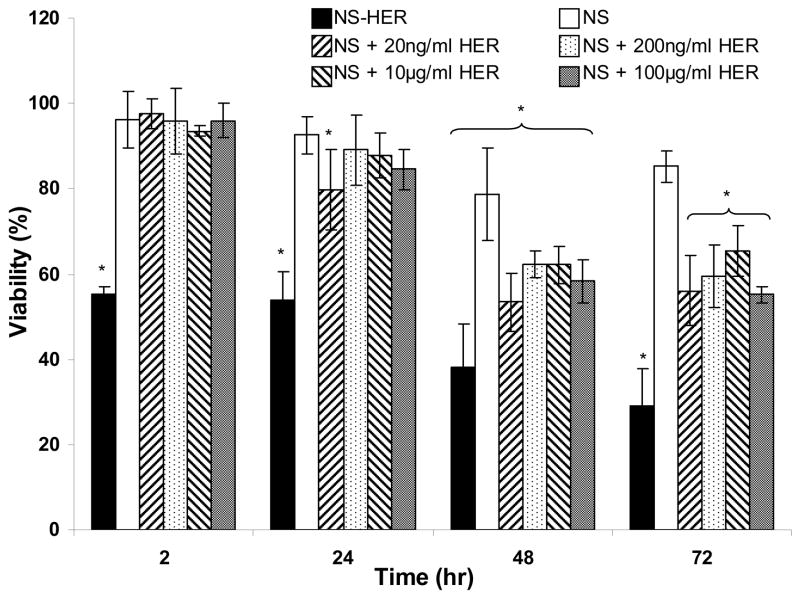
Figure 5.
Cytotoxicities of NS-HER and NS with various concentrations of herceptin, as measured by the viabilities of SK-Br-3 cells grown in media containing 0.2 mg/ml of these nanospheres relative to the non-toxic control. Results are represented as mean ± standard deviation. “*” denotes statistical differences (P < 0.05) compared to the control experiment.

