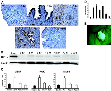
Figure 2.
A: Immunohistochemistry staining of endometriosis-like lesions with anti-HIF-1α antibody. Counterstaining with hematoxylin. Increasing immunoreactivity (brown color) in glandular epithelial and stromal cells (arrows) over time (0, 1, 3, and 6 hours). Control (6 hours) stained with nonspecific rabbit IgG antibody. B: Western blot of endometriotic tissue stained with ant-HIF-1α antibody from different time points after transplantation. Loading control of same Western blot stained with anti-β-actin antibody. C: Real-time RT PCR of endometriosis-like lesions at different time points using primers for HIF-1α-dependent genes (VEGF, PGK, and Glut-1). Bars indicate SEM. D: Semiquantitative measurement of Western blot for HIF-1α as seen in B. Error bars indicate SEM. E: Endometriosis-like lesion from a green fluorescent protein+/+ mouse on the peritoneal wall of a wild-type recipient. Typical growth of blood vessels into the endometriosis-like lesion.

