Figure 1.
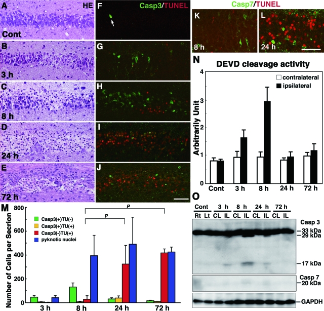
Activation of caspases and TUNEL staining in the hippocampus of wild-type neonatal mice after H/I injury. A–J: Histological sections of the left hippocampus of an untreated control (Cont) mouse at P7 (A, F) and ipsilateral hippocampus obtained 3 (B, G), 8 (C, H), 24 (D, I), and 72 hours (E, J) after H/I injury and stained with H&E (A–E) or for TUNEL and the active form of caspase-3 (F–J). An arrow in F shows a cleaved caspase-3-positive neuron undergoing programmed cell death. K and L: Double staining for TUNEL and the cleaved form of caspase-7 in the ipsilateral hippocampus at 8 (K) and 24 (L) hours after H/I injury. M: Number of neurons with pyknotic nuclei stained by H&E and the number of neurons positive for the active form of caspase-3 (Casp3), TUNEL (TU), or both in the pyramidal layers per hippocampal section at 3, 8, 24, and 72 hours after H/I injury. Counts were taken from three sections per animal, and the final value presented is the mean ± SD for three animals. P < 0.001. N: Proteolytic activity of the DEVDase for caspases-3 and -7 in the ipsilateral and contralateral hippocampi 3, 8, 24, and 72 hours after H/I injury. The activity in the nontreated hippocampus was used as a control. The final value presented is the mean ± SD for three animals. O: The cleavage of caspases-3 and -7 in the contralateral (CL) and ipsilateral (IL) hippocampi of wild-type neonatal mouse brains at 3, 8, 24, and 72 hours after H/I injury. Hippocampal tissues of the right (Rt) and left (Lt) sides from untreated neonatal mice were used as controls. Scale bars = 50 μm.

