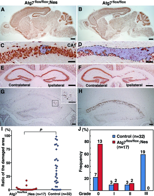
Figure 6.
Atg7 deficiency prevents neuron death in the hippocampus of neonatal mouse brains 3 days after H/I injury. A–H: Saggital (A–D) and frontal (E–H) sections of the Atg7-deficient mouse brains (Atg7flox/flox; Nes) in the CNS (A, C, E, G) and its littermate (Atg7flox/flox) (B, D, F, H) 3 days after H/I injury. NeuN (A–F) and TUNEL (G, H) staining. Damaged areas in the hippocampus of A and B are shown in C and D and encircled by broken lines. I and J: Evaluation of damaged areas. I: Ratios (%) of damaged areas to the total areas in the hippocampal layers of Atg7-deficient (n = 17) and littermate control (n = 32) mouse brains 3 days after H/I injury. P < 0.001 (Mann-Whitney U-test for the comparison of the median values between the two groups). J: Incidence of hippocampal damage 3 days after H/I injury. The severity of the H/I injury was graded according to the number of TUNEL-positive neurons in the pyramidal layers of the hippocampus persection (less than 10 for grade 0, 11 to 100 for grade I, 101 to 200 for grade II, and more than 201 for grade III). The percentage of mice classified into each grade was determined from 17 Atg7 flox/flox; nestin-Cre (Atg7flox/flox; Nes), and 32 control littermate (Atg7flox/flox) mice. The number of mice belonging to each grade was described in the top of each bar. Scale bars: 500 μm (A, B, E, F); 50 μm (C, D); 100 μm (G, H, inset of G).

