Figure 4.
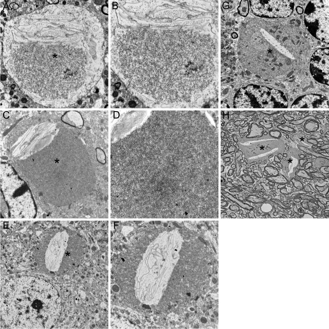
Transmission EM reveals tubulovesicular membranes and other material accumulating within spheroids. A–F: Images from transmission EM, performed on sections from striatum of 8-month-old KO and WT mice, illustrate ultrastructural features of spheroids. B, D, and F are higher magnification images of A, C, and E, respectively. Spheroids (asterisks) were typically surrounded by an intact membrane. Common to all spheroids was the presence of accumulating membranes. Tubulovesicular membranes were abundant and interrupted or bordered by clefts containing sheets of membranes, often in stacked or whorled structures. These structural features of spheroids strongly resemble those reported in human INAD. Transmission EM in the cerebellar granule layer (G) and molecular layer (H) also indicates accumulation of tubulovesicular membranes in spheroids. Magnifications: ×6400 (A, C, F), ×8400 (B), ×12,800 (D), ×3200 (E), ×4200 (G), ×1280 (H).

