Figure 3.
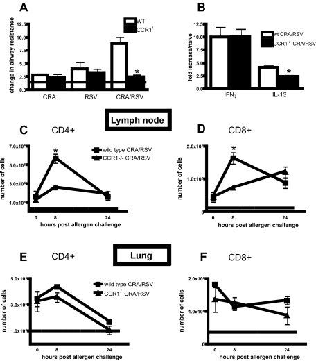
CCR1−/− mice have reduced AHR and T-cell recruitment during viral exacerbation of allergic airway disease. A: AHR of CCR1−/− and wild-type mice was assessed 24 hours after antigen challenge or 6 days after RSV infection. The dashed line represents AHR of naïve mice. *P = 0.0022. B: The amount of transcript for IL-13 and IFN-γ was assessed using whole lung mRNA by real-time PCR. *P = 0.024. C–F: The number of CD4+ and CD8+ T cells in the lungs and lymph nodes of exacerbated wild-type and CCR1−/− mice at 0, 8, and 24 hours after antigen challenge. The dashed line represents the number of T cells found in the lungs or lymph nodes of naïve mice. *P < 0.01. n = five mice/group/experiment, data are pooled from two experiments.

