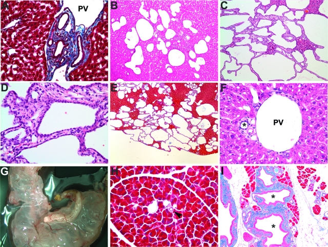
Figure 2.
Histological analysis of Pkhd1del4/del4 livers and pancreas. A: Mallory trichrome staining of 2-week-old Pkhd1del4/del4 liver. B–D: H&E staining of Pkhd1del4/del4 liver at 3 months (B) and at 12 months (C, D). E: Mallory trichrome staining of Pkhd1del4/del4 liver at 12 months. F: H&E staining of 12-month-old wild-type bile ducts indicated by the asterisk. G: Grossly cystic pancreas from a 3-month-old Pkhd1del4/del4 animal. H and I: Mallory trichrome staining of the pancreas from 6-month-old wild-type (H) and Pkhd1del4/del4 (I) mice showing pancreatic ductal dilatation in mutant mice. The pancreatic ducts are highlighted by an arrowhead in H and by an asterisk in I. PV, portal vein. Original magnifications: ×400 (A, D, F); ×100 (B, C, H, I); ×40 (E).

