Figure 6.
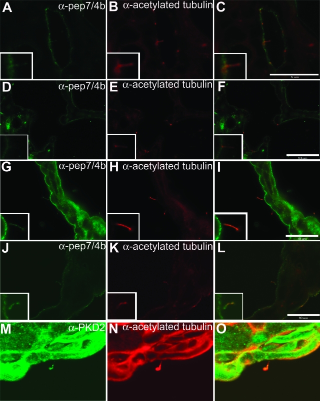
Trafficking of fibrocystin and PC2 in cyst-lining bile duct epithelia of ADPKD and Pkhd1del4/del4 mouse models. A-C: Fibrocystin is expressed at the apical membranes and cilia of wild-type hepatic bile ducts. D–F: In Pkhd1del4/del4 mice, mutant fibrocystin can still be detected in the apical membranes and cilia of cyst-lining epithelia in the liver. G–L: Immunohistochemical analysis of wild-type fibrocystin expression in cyst-lining epithelia of Pkd1 cystic kidney (G–I) and Pkd2 cystic liver (J–L) reveals normal-appearing expression of fibrocystin in apical membranes and cilia in these cystic tissues. Cilia were identified by co-staining with acetylated α-tubulin (B, E, H, K). PC2 is expressed normally in cilia of cyst-lining biliary epithelia of Pkhd1del4/del4 mice. Original magnification, ×240 (M–O).

