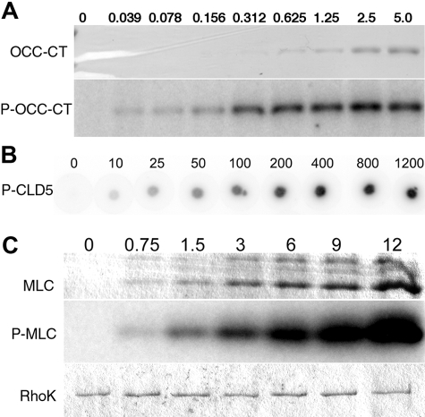
Figure 1.
Dose-dependent analysis of phosphorylation of CLD5-CT peptide, MLC, and OCC-CT by GST-RhoK. A and C: Increasing doses of OCC-CT (0 to 5 μmol/L, A) or MLC (0 to 12 μmol/L, C) were incubated with [γ-32P] ATP and purified GST-RhoK and separated on SDS-PAGE, followed by R-250 Coomassie brilliant blue staining (CBB, top) and autoradiogram (middle) of OCC-CT (A) or MLC (C). B: Increasing doses of CLD5-CT (0 to 1200 μmol/L, B) or were phosphorylated by GST-RhoK, and spotted onto phosphocellulose membrane. After extensive washing, the 32PO4 incorporation was visualized by Typhoon Phosphoimager. C: Bottom shows the CBB-stained GST-RhoK on the same gel.

