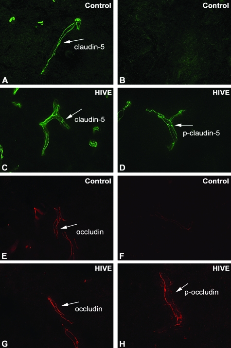
Figure 10.
Detection of OCC and CLD5 phosphorylation in brain tissues of hu-PBL-NOD/SCID HIVE mice. A–H: Ten-μm-thick frozen sections of mouse brains were prepared at 14 days after sham injection (A, B, E, F) or intracerebral injection of HIV-1-infected MDM (C, D, G, H). The sections were stained with anti-CLD5 (A, C), anti-CLD5 pT207 (B, D), anti-OCC (E, G), or anti-OCC pT382 antibodies (F, H) and fluorescence-conjugated secondary antibodies. A–H: Oil immersion. Original magnifications, ×1000.

