Figure 12.
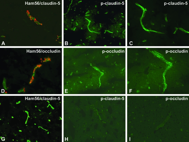
Expression of phosphorylated TJ proteins in human brain tissues with HIVE. Ten-μm-thick frozen sections of human brains with HIVE (A–F) or controls (G–I) were cut. The sections were double immunostained with Ham56 (macrophage marker) and anti-CLD5 (A, G) or OCC (D), anti-CLD5 pT207 (B, C, H), or anti-OCC pS507 antibodies (E, F, I) and fluorescence-conjugated secondary antibodies. CLD5 (green, A) and OCC (green, D) showed fragmented staining in areas containing inflammatory cells (red, Ham56 monocyte/macrophage marker). Antibodies to p-CLD5 (pT207, B and C) and p-OCC (pS507, E and F) highlighted microvessels (arrows). Control brains demonstrated contiguous CLD5 immunostaining (green) and few perivascular monocytes/macrophages (red) (CLD5, G), whereas phosphospecific antibodies to TJ proteins produced no staining (OCC pS507, H; CLD5 pT207, I). Original magnifications: ×200 (A, B, D, E, G, H, I); ×400 (C, F).

