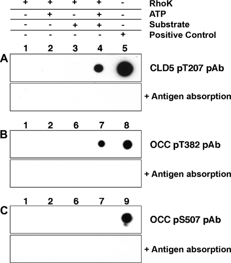
Figure 6.
Detection of CLD5 and OCC phosphorylation by specific anti-phosphopeptide antibodies. A–C: Top: CLD5 cytoplasmic sequence peptide (A, 500 ng per spot) or OCC cytoplasmic protein fragment (B and C, 2 μg per spot) were incubated with GST-RhoK (200 ng/μl) overnight at 30°C, and spotted onto polyvinylidene difluoride membrane for dot-blotting using anti-CLD5 pT207 (A), anti-OCC pT382 (B), or anti-OCC pS507 antibody (C). Each antibody was preincubated with 10 μg/ml of corresponding antigen phosphopeptide before the immunoblotting for antigen absorption (bottom, A–C). Lane 1, Two hundred ng of RhoK without ATP; lane 2, 200 ng of RhoK with 1 mmol/L ATP; lane 3, 500 ng of CLD5 peptide with 200 ng of RhoK without ATP; lane 4, 500 ng of CLD5 peptide with 200 ng of RhoK and 1 mmol/L ATP; lane 5, 500 ng of CLD5 pT207 phosphopeptide; lane 6, 2 μg of OCC-CT with 200 ng of RhoK without ATP; lane 7, 2 μg of OCC-CT with 200 ng of RhoK and 1 mmol/L ATP; lane 8, 500 ng of OCC pT382 phosphopeptide; lane 9, 500 ng of OCC pS507 phosphopeptide.

