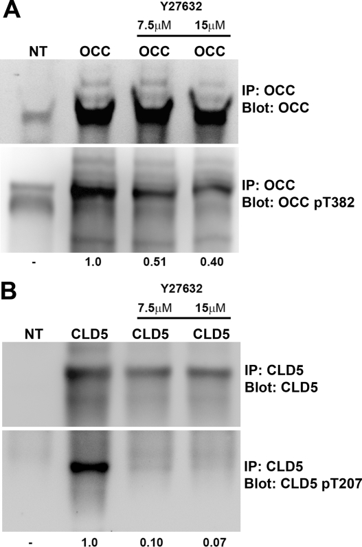
Figure 7.
Detection of OCC and CLD5 phosphorylation in COS-7 cells. COS-7 cells were transfected with full-length wild-type mouse OCC or CLD5 mammalian expression vector, and treated or untreated with RhoK inhibitor Y27632 (7.5 or 15 μmol/L) for 16 hours. The plasma membrane samples were lysed in 1% Trion X-100 and subjected to immunoprecipitation using 20 μg of anti-OCC (A), or CLD5 mouse monoclonal antibodies (B) plus protein A/G Plus-Sepharose. Immunoblotting for OCC and p-OCC (pT382, A) or CLD5 and p-CLD5 (pT207, B) was performed on immunoprecipitated samples using specific rabbit polyclonal antibodies. IP and blot (A and B) indicate specific molecules for immunoprecipitation and immunoblotting, respectively. A and B: NT; no transfection; OCC, transfected recombinant mouse OCC; Cld-5, recombinant mouse CLD5. The number at the bottom indicates changes in arbitrary unit of the immunoreactive band intensity of p-OCC (pT382, A) or p-CLD5 (pT207, B) after Y27632 treatment.

