Figure 8.
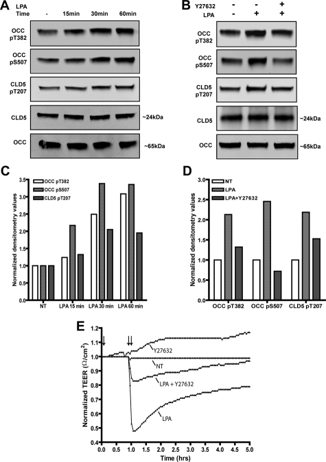
Detection of LPA-induced OCC and CLD5 phosphorylation and TEER reduction in mouse BMVECs. Western blots were performed using novel phosphospecific antibodies, OCC pT382, OCC pS507, and CLD5 pT207 in mouse brain endothelial cells; nonphoshospecific antibodies recognizing total OCC and total claudin5 are also indicated. A and C: Cells were cultured under reduced serum conditions (1% FBS) for 24 hours and then stimulated with 10 μmol/L LPA for the indicated time points; or B and D: pretreated with 2 μmol/L Y27632 for 1 hour followed by stimulation with 10 μmol/L LPA for 30 minutes. Corresponding densitometry analysis of the Western blots is shown below (C, D), the densitometry values represent the ratio of phosphorylated to unphosphorylated as compared with the nontreated (NT) condition. E: Endothelial cell monolayers were formed on ECIS electrode culture slides (described in Materials and Methods) and real time measurements of TEER were acquired. The measurements shown represent recordings acquired at 3-minute intervals at the parameters described in the Materials and Methods. The single arrow designates the pretreatment (1 hour) with 2 μmol/L of Y27632 with or without co-incubation of 10 μmol/L LPA as shown by the double arrow. The data are presented as normalized resistance for each condition, which is the resistance measured after treatment over the resistance acquired before treatment introduction (typically ∼2600 Ω/cm2). Representative data of three independent experiments are shown.

