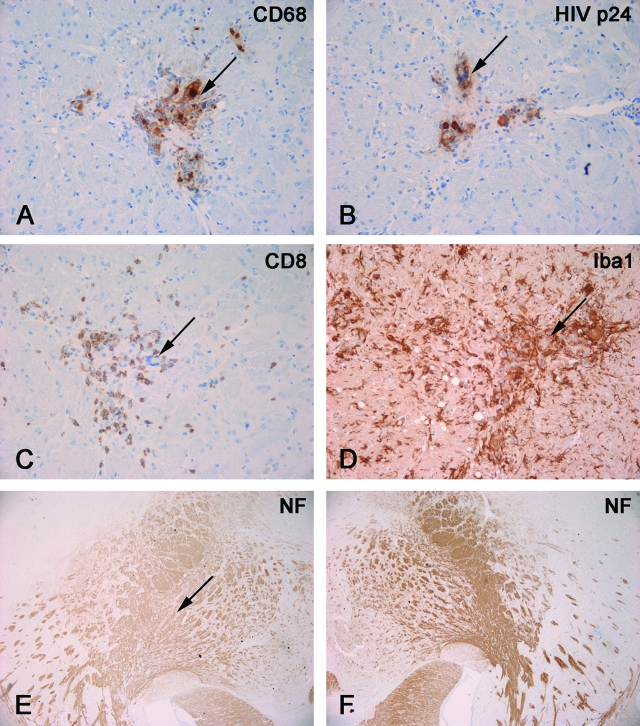
Figure 9.
Neuropathological features of HIVE in hu-PBL-NOD/SCID HIVE mice. Mice were reconstituted with human PBL and sacrificed 2 weeks after injection of HIV-1-infected MDM in brains. Serial mouse brain sections stained with antibodies to human CD68 (A), HIV-p24 (B), human CD8 (C), Iba1 (D), or NF (E, F). Primary Abs were detected by Vectastain Elite kit with DAB as a substrate. A and B: CD68+ human MDM (arrow, A) in the basal ganglia were HIV-1 p24+ (arrow, B). C: CD8+ lymphocyte infiltrated in the areas containing virus-infected macrophages (arrow). D: These areas featured prominent microglial reaction (Iba1+, arrow indicates location of MDM). E and F: Decreased staining for neuronal marker (NF) was found in injected hemisphere (arrow indicates area of injection, E) as compared to contralateral (noninjected, F). Original magnifications: ×200 (A–D); ×40 (E, F).

