Figure 1.
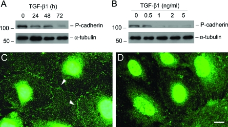
TGF-β1 inhibits P-cadherin expression in podocytes. A and B: Western blot analyses show that TGF-β1 inhibited P-cadherin protein expression in a time- and dose-dependent manner. Mouse podocytes were incubated with either the same concentration of TGF-β1 (2 ng/ml) for various periods of time as indicated (A), or increasing amounts of TGF-β1 for 72 hours (B). Cell lysates were immunoblotted with antibodies against P-cadherin and α-tubulin, respectively. C and D: Immunofluorescence staining shows the localization of P-cadherin in control (C) or TGF-β1-treated podocytes (D). Arrowheads indicate the positive P-cadherin staining. Scale bar = 10 μm.

