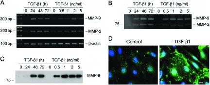
Figure 6.
TGF-β1 induces MMP-9 production and secretion by podocytes. A: RT-PCR analysis demonstrates that TGF-β1 induced MMP-9 and MMP-2 mRNA expression in podocytes. Mouse podocytes were incubated with TGF-β1 (2 ng/ml) for various periods of time or with increasing amounts of TGF-β1 for 72 hours. B: Zymographic analysis of the conditioned media derived from podocytes treated without (control) or with TGF-β1 as indicated. Samples equalized for protein content were separated on a 10% sodium dodecyl sulfate-polyacrylamide gel containing 0.1% gelatin. The locations of bands corresponding to MMP-9 and MMP-2 are indicated. C: Western blot analysis of the conditioned media from podocytes treated without (control) or with TGF-β1 as indicated. D: Immunofluorescence staining demonstrates the distribution of MMP-9 in the control and TGF-β1-treated podocytes. Scale bar = 10 μm.

